Inflammation
Scalp burning, tenderness, itching, or red or discoloured patches.
Scarring alopecia, also called cicatricial alopecia, occurs when hair follicles are permanently damaged and replaced by scar tissue. In many forms, ongoing inflammation causes this damage. Some cases cause itching, burning, tenderness, scale or pustules; others progress quietly. Treatment is directed at stopping further destruction, not promising regrowth in completely scarred areas.
Consultation ₹800 · Pre-booking required · No advance booking payment
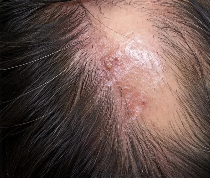
These clues help distinguish one kind of hair loss from another—but images and symptoms alone cannot confirm a diagnosis.
Scalp burning, tenderness, itching, or red or discoloured patches.
Scales or redness concentrated around emerging hairs.
Shiny areas with reduced or absent follicular openings.
An expanding patch or front hairline moving backwards.
In male and female pattern hair loss the follicles miniaturise, and in alopecia areata they usually remain intact. In scarring alopecia, inflammatory injury can replace a follicle with fibrous tissue. Once a follicle is destroyed, medication generally cannot make hair grow from that site again.
Important forms include lichen planopilaris, frontal fibrosing alopecia, discoid lupus of the scalp, central centrifugal cicatricial alopecia and some destructive folliculitis disorders. Their treatments differ, so a precise diagnosis matters.
Scarring alopecia is a group of conditions, not a single diagnosis. The pattern of hair loss, inflammation and symptoms helps guide assessment and treatment.
Irregular patches of hair loss with redness or scale around hair follicles; itching, burning or tenderness may occur.
Gradual recession of the frontal or temple hairline, sometimes accompanied by thinning or loss of the eyebrows.
Inflamed, scaly patches that may leave lighter or darker skin changes and permanent scarring hair loss.
Repeated scalp pustules or crusting, sometimes with several hairs emerging together from one follicular opening (tufting).
Painful, swollen scalp nodules or boggy areas that can discharge pus and eventually cause scarring.
Hair thinning and loss that often starts around the crown and gradually spreads outward, sometimes with itching or tenderness.
These appearances can overlap. A dermatologist may use trichoscopy and, when indicated, a scalp biopsy to identify the cause before selecting treatment.
History and scalp examination guide the need for trichoscopy, a biopsy, or targeted tests (such as bacterial or fungal testing when infection is suspected). Not everyone needs every investigation.
Look for loss of follicular openings, perifollicular erythema/scale, pustules, changes at the front hairline and eyebrow involvement.
Identify active inflammatory areas and guide selection of a biopsy site.
A biopsy from an actively inflamed area, often near the edge rather than the fully scarred centre, may help identify the subtype and guide treatment.
If a biopsy is needed, a small sample of scalp skin (often around 4 mm) is usually taken under local anaesthesia from an active area near the edge of hair loss rather than the middle of an old scar. A stitch may be required, and a small mark or scar can remain. The tissue is examined under a microscope; results are interpreted together with scalp examination and trichoscopy to guide diagnosis and treatment. Not every patient needs a biopsy.
Management depends on the actual condition, its severity, age, medical history and your goals. Procedures are not a replacement for treating the underlying cause.
The main goal is controlling inflammation and preventing additional permanent hair loss. Early treatment is especially important.
Depending on the subtype, treatment may include topical/injected corticosteroids, other anti-inflammatory agents or prescription systemic medicines with appropriate monitoring.
Assess pustules or possible infection, review damaging hair-care practices where relevant, and manage associated scalp symptoms. Not every scarring disorder is autoimmune.
Completely scarred follicles are unlikely to regrow. Minoxidil may help surviving neighbouring follicles in selected patients but does not treat active scarring inflammation by itself; PRP/GFC is not a substitute for anti-inflammatory care.
The aim is to stop additional follicle damage. Follow-up checks whether inflammation is settling and hair loss has stabilised; visible regrowth is not expected in areas where follicles have already scarred.
Avoid delay: investigate active inflammatory hair loss promptly.
Monitor symptoms, scalp inflammation, progression and treatment adverse effects.
Aim for disease inactivity; remaining hair can often be protected better than already scarred sites restored.
Comparable photographs of the same scalp areas help show whether patches or the hairline are changing over time.
Reduction in redness, scale, pustules and other signs of active inflammation can suggest better disease control.
Itching, burning, tenderness, new patches and hairline changes are reviewed. Some disease can progress without symptoms.
If prescription medicines are used, the dermatologist also checks tolerability, possible adverse effects and any necessary monitoring tests.
Clear answers without unrealistic promises or one-size-fits-all treatments.
Do not begin or discontinue prescription treatments solely from a webpage. A dermatologist can identify overlapping disorders and discuss benefits, contraindications and follow-up. All clinical images on this page are AI-generated illustrations—not photographs of patients or treatment results.
Dr Neeraj Garg · MBBS (IMS-BHU), MD (IMS-BHU)
Maheshwari Hospital, Dalanwala, Dehradun · Consultation ₹800